Adult cardiomyocyte renewal, reprogramming, tissue replacement, fibrosis, metabolism, and the evidence required to call an intervention regenerative.
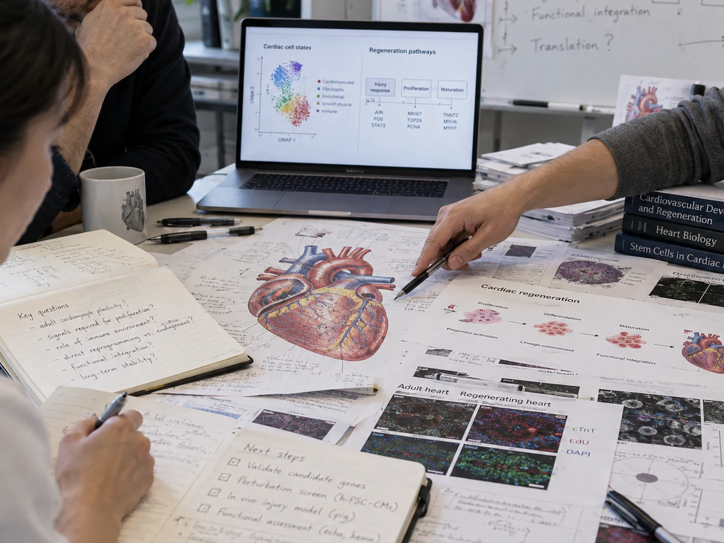
Adult mammalian cardiomyocytes are highly specialized for force generation and electrical coupling, but that mature state is linked to strong barriers to cell-cycle re-entry. Postnatal maturation changes chromatin, metabolism, sarcomere organization, centrosome biology, ploidy, extracellular matrix, and oxygen handling. After infarction, rapid inflammatory and fibroblast responses stabilize the ventricle but also favor scar formation over remuscularization.
Many adult cardiomyocytes can enter partial cell-cycle programs without completing cytokinesis. A convincing regenerative claim must distinguish DNA synthesis, karyokinesis, and actual generation of new cardiomyocytes.
The mature sarcomere, metabolic program, electrophysiologic specialization, and nuclear architecture support function but create structural and transcriptional resistance to proliferation.
Fibroblast activation and matrix deposition preserve wall integrity after injury. Regeneration therefore cannot be framed as “remove fibrosis”; the challenge is to coordinate mechanical stability, vascular repair, inflammation, and tissue replacement.
Endothelial, immune, fibroblast, neural, extracellular-matrix, and metabolic cues influence whether a cardiomyocyte remains mature, becomes stressed, dedifferentiates, or re-enters a proliferative program.
| Axis | Biological role | Regenerative opportunity | Main risk |
|---|---|---|---|
| Cyclin / CDK network | Controls G1/S and G2/M progression. | Re-open a normally quiescent cardiomyocyte cell cycle. | Polyploidy, incomplete cytokinesis, dysregulated proliferation. |
| Hippo–YAP | Links tissue architecture and growth control to transcription. | Increase pro-proliferative and pro-survival programs. | Loss of temporal control, abnormal growth, non-cardiomyocyte effects. |
| Metabolism / oxygen | Postnatal shift toward oxidative metabolism accompanies maturation. | Transiently reproduce a more permissive metabolic state. | Energetic impairment or maladaptive stress responses. |
| Chromatin / epigenetics | Locks mature identity and suppresses cell-cycle programs. | Lower barriers to re-entry or lineage conversion. | Broad, poorly specific transcriptional effects. |
| ECM / mechanics | Controls force transfer, stiffness sensing, and fibroblast activation. | Build a pro-renewal mechanical niche while preserving wall strength. | Rupture, maladaptive remodeling, arrhythmogenic heterogeneity. |
| Immune / vascular niche | Coordinates injury clearance, angiogenesis, and remodeling. | Shift from persistent inflammation toward constructive repair. | Immunosuppression, excess angiogenesis, off-target tissue effects. |
Stimulate pre-existing cardiomyocytes to re-enter and complete the cell cycle. The key challenge is producing enough new cells while preserving electrical and mechanical function.
Convert fibroblasts toward induced cardiomyocyte-like states using transcription factors, RNAs, small molecules, or combinations. Human-cell efficiency and maturation remain major barriers.
Engraft cardiomyocytes derived from pluripotent stem cells or engineered tissue. Survival, vascularization, immune compatibility, maturation, and arrhythmia risk dominate translation.
Use biomaterials, paracrine signals, extracellular vesicles, or immune modulation to improve survival, vascularization, matrix organization, and endogenous repair.
Single proliferation markers are not enough. Strong evidence needs lineage-aware cell counting, cytokinesis-sensitive assays, independent functional measurements, arrhythmia assessment, and durability over time.
The regeneration biology page is the scientific substrate for target discovery, VB-101 candidate ranking, VB-204 tissue design, and HeartTwin hypothesis testing. It does not imply that the current stack has demonstrated myocardial regeneration.